

 Tiếng Việt
 Русский
Sinus lift surgery (maxillary sinus floor elevation) in dental implantology is an essential regenerative procedure designed to increase vertical alveolar bone volume in the posterior maxilla, establishing a stable foundation for osseointegration. Typically indicated when residual subantral bone height is insufficient, this technique ensures predictable, long-term implant survival. Explore the clinical workflows, indications, and benefits of sinus augmentation in the guide below!
What Is a Sinus Lift in Dental Implantology?
Sinus lift surgery in dental implant placement is a specialized pre-prosthetic surgical procedure performed when the posterior maxillary alveolar ridge lacks sufficient vertical height and bone volume—due to post-extraction resorption or sinus pneumatization—to securely anchor an endosseous implant fixture.
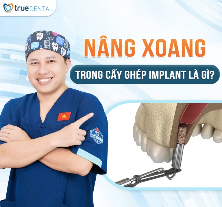
Following prolonged loss of maxillary posterior teeth—particularly the first and second premolars and molars (teeth #4, #5, #6, and #7)—natural ridge resorption combined with pneumatization (inferior expansion) of the maxillary sinus reduces residual bone thickness. Sinus floor elevation carefully lifts the Schneiderian membrane (sinus lining) upward and packs bone graft material into the subantral space, creating sufficient bone height for safe, durable implant placement.
Depending on residual alveolar bone height and sinus anatomy, specialists utilize one of two primary surgical approaches: a closed (transcrestal) sinus lift or an open (lateral window) sinus lift.
Open Sinus Lift (Lateral Window Technique)
An open sinus lift is a more extensive surgical augmentation indicated when residual subantral bone height is less than 4 mm. In this approach, the surgeon elevates a mucoperiosteal flap and creates a small lateral bony window on the buccal wall of the maxillary sinus to access the Schneiderian membrane directly.
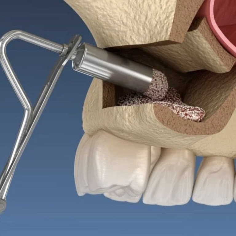
After gently elevating the sinus membrane, particulate bone graft material is placed into the compartment to regenerate substantial vertical bone height. Because it involves greater surgical access, recovery takes slightly longer than a closed approach, yet it delivers superior predictability in cases of severe maxillary atrophy.
Closed Sinus Lift (Transcrestal / Osteotome Technique)
A closed sinus lift is a minimally invasive technique typically indicated when residual vertical bone height measures between 4 and 6 mm. The specialist accesses the sinus floor directly through the crestal implant osteotomy site, using specialized osteotomes or hydraulic lifters to elevate the sinus membrane gently.
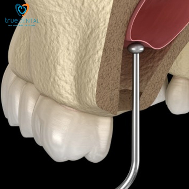
Alloplastic, xenograft, or autogenous bone graft material is then introduced through the osteotomy into the elevated space. This transcrestal approach offers faster healing, minimal post-operative discomfort, and is ideal for mild-to-moderate vertical bone deficiencies.
When Is Sinus Augmentation Indicated?
Sinus augmentation is indicated whenever residual alveolar bone height or trabecular bone density in the posterior maxilla is insufficient to achieve primary implant stability and long-term biomechanical support. It is an indispensable preparatory step for patients with long-standing posterior edentulism or advanced sinus pneumatization.
Performing sinus floor elevation not only restores maxillary ridge architecture but also ensures balanced occlusal load distribution and long-term prosthetic success. If you are considering upper posterior implants and suspect insufficient bone height, consult an implant specialist for a 3D CBCT evaluation.
Is Sinus Lift Bone Grafting Mandatory for Implant Placement?
Sinus lift bone grafting during dental implant surgery is mandatory when the posterior maxilla lacks adequate vertical bone height or density to house an implant fixture safely without perforating the sinus cavity—a common scenario in long-term tooth loss.
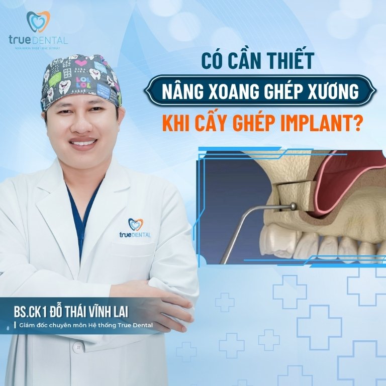
By regenerating lost vertical bone volume, sinus augmentation ensures the fixture is anchored securely in dense bone capable of withstanding heavy posterior masticatory forces. Attempting implant placement without necessary sinus grafting significantly increases the risk of sinus membrane perforation, chronic sinusitis, failed osseointegration, or fixture loss.
Determining the need for sinus augmentation relies on precise preoperative diagnostics. Clinical examination paired with 3D Cone Beam CT imaging enables your dental specialist to measure exact subantral bone dimensions and formulate a safe, predictable treatment plan.
Indications and Contraindications for Sinus Lift Surgery
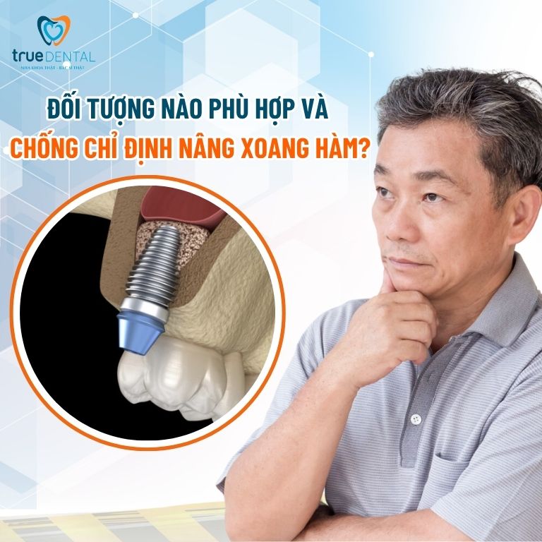
While maxillary sinus floor elevation offers high clinical predictability, proper patient selection is essential to avoid surgical complications. Key indications and contraindications include:
Clinical Indications
- Patients exhibiting posterior maxillary alveolar ridge resorption or sinus pneumatization resulting in insufficient vertical bone height for standard implant lengths.
- Individuals with long-standing posterior tooth loss leading to progressive bone atrophy.
- Patients requiring enhanced subantral bone volume to ensure long-term biomechanical stability of implant-supported crowns or bridges.
- Candidates free of active maxillary sinus pathology and in stable systemic health to undergo minor oral surgery.
Contraindications
- Uncontrolled systemic conditions such as poorly controlled diabetes mellitus, active malignancies, severe bleeding disorders, or immunocompromised states.
- Active maxillary sinus pathology, including acute or chronic rhinosinusitis, large sinus polyps/cysts, or odontogenic infections in the posterior maxilla.
- Pregnant or lactating women, for whom elective surgical interventions are deferred.
- Patients with severely compromised bone metabolism or local anatomical defects precluding predictable graft consolidation.
Candidacy for sinus augmentation must always be established by an experienced implantologist following comprehensive clinical and 3D radiographic evaluation to ensure safety and optimal outcomes.
Comparison of Closed vs. Open Sinus Lift Techniques
Contemporary implantology utilizes two primary sinus elevation techniques—closed (transcrestal) and open (lateral window)—each tailored to specific residual bone heights and anatomical requirements:
| Comparison Criteria | Closed Sinus Lift (Transcrestal) | Open Sinus Lift (Lateral Window) |
| Surgical Access | Via the crestal implant osteotomy site | Via a lateral bony window prepared on the buccal sinus wall |
| Clinical Indications | Residual bone height of 4–6 mm, flat sinus floor, mild-to-moderate resorption | Severe posterior atrophy, residual bone height less than 4 mm, complex sinus septa |
| Invasiveness | Minimally invasive | More invasive, requiring lateral mucoperiosteal flap elevation |
| Recovery Timeline | Faster healing with minimal pain and swelling | Longer recovery with moderate swelling requiring close monitoring |
| Simultaneous Implant Placement | Frequently performed simultaneously in a single visit | Usually staged (waiting 4–6 months for graft maturation before placement) |
Clinical Workflow for Sinus Lift Surgery
Depending on the degree of alveolar ridge resorption and sinus floor anatomy, your specialist will execute either the closed or open sinus lift protocol:
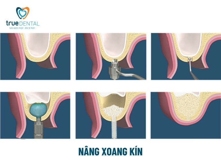
Closed (Transcrestal) Sinus Lift Protocol
- Clinical Examination & 3D Diagnostics: The clinician evaluates oral health and analyzes 3D Cone Beam CT scans to measure exact residual subantral bone height and sinus floor morphology.
- Crestal Osteotomy Preparation: Under local anesthesia, a narrow 3–4 mm crestal channel is prepared at the edentulous site just short of the sinus floor.
- Schneiderian Membrane Elevation: Specialized osteotomes or hydraulic sinus lifters are used to gently fracture the sinus floor and elevate the Schneiderian membrane upward, creating a subantral compartment.
- Bone Graft Placement: Particulate bone graft material is carefully condensed through the osteotomy beneath the elevated membrane to achieve the desired vertical height.
- Simultaneous Implant Placement & Suturing: When adequate primary stability is achieved, the implant fixture is inserted immediately during the same procedure, followed by tension-free closure with resorbable sutures.
- Post-Operative Radiographic Verification: A control radiograph confirms accurate 3D fixture alignment and containment of the bone graft beneath an intact sinus membrane.
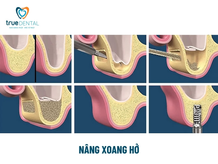
Open (Lateral Window) Sinus Lift Protocol
- Diagnostic Evaluation & Surgical Planning: The specialist evaluates 3D CBCT scans to map sinus septa, lateral wall thickness, and the required graft volume for severe vertical bone deficiency.
- Flap Elevation & Lateral Window Creation: A mucoperiosteal flap is reflected, and a precision lateral window is prepared in the buccal sinus wall using piezosurgery or diamond burs to expose the Schneiderian membrane.
- Membrane Detachment & Elevation: Delicate sinus curettes are used to carefully detach and elevate the Schneiderian membrane superiorly and medially without perforation.
- Bone Grafting & Membrane Coverage: Bone graft biomaterial is packed into the newly created subantral space, and a resorbable collagen barrier membrane is placed over the lateral window to protect graft consolidation.
- Primary Closure & Graft Maturation: The soft tissue flap is repositioned and sutured closed, followed by a 4- to 6-month healing interval for bone remodeling and vascularization.
- Staged Implant Placement: Once radiographic evaluation confirms dense bone maturation, the implant fixture is placed with high primary stability.
Can an Implant Be Placed Simultaneously with a Sinus Lift?
Whether an implant fixture can be placed simultaneously during sinus lift surgery depends on the chosen elevation technique and the residual native bone available to achieve primary mechanical stability.
In a closed (transcrestal) sinus lift—where 4 to 6 mm of dense native bone remains—simultaneous implant placement is routinely performed. Immediately after elevating the membrane and placing the bone graft, the specialist inserts the implant fixture in the same surgical visit, condensing total treatment time and minimizing surgical appointments.
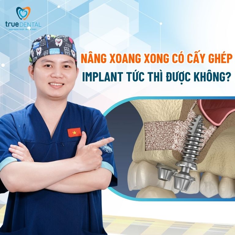
Conversely, in an open (lateral window) sinus lift—indicated when residual bone is less than 4 mm—primary stability cannot always be guaranteed immediately. In these cases, clinicians follow a staged approach, waiting 4 to 6 months for the bone graft to consolidate and mineralize into mature bone before placing the implant fixture.
Ultimately, the decision between simultaneous and staged placement must be made by an experienced implantologist based on 3D CBCT diagnostics to ensure maximum clinical safety and osseointegration success.
Post-Operative Care Guidelines After Sinus Lift Surgery
Strict adherence to post-operative care protocols following maxillary sinus augmentation promotes uneventful graft healing and prevents sinus membrane complications:
- Prioritize Rest & Head Elevation: Avoid strenuous physical exertion for at least 1–2 weeks. Sleep with your head elevated on extra pillows during the first few nights to minimize facial edema and sinus pressure.
- Protect Sinus Pressure & Surgical Site: Never touch the wound with fingers or tongue. Avoid forceful nose blowing, spitting, or drinking through straws, and sneeze with your mouth open to prevent sudden pressure spikes across the sinus membrane.
- Follow a Soft, Nutrient-Rich Diet: Consume cool-to-lukewarm soft foods during initial recovery, avoiding hard, crunchy, spicy, or acidic items. Stay well-hydrated and maintain adequate vitamin intake to support tissue repair.
- Avoid Barometric Pressure Changes: Refrain from air travel, scuba diving, or wind-instrument playing during the initial healing phase as advised by your surgeon.
- Attend Scheduled Follow-Up Visits: Keep all post-operative appointments so your specialist can monitor soft tissue closure and radiographic graft consolidation.
- Take Prescribed Medications Strictly as Directed: Complete prescribed antibiotics, analgesics, and nasal decongestants/saline sprays to prevent sinus infection and ensure comfortable recovery.
READ MORE:
Maxillary sinus floor elevation is a highly predictable regenerative solution for patients with posterior maxillary bone loss and sinus pneumatization. Choosing between a closed or open sinus lift depends on your individual bone dimensions and specialist evaluation. Contact True Dental today for a comprehensive 3D consultation and personalized treatment plan!

Sức Khỏe Răng Miệng
How long does it take to complete the process of getting dental implants?
Tooth replacement affects far more than just smile...
Sức Khỏe Răng Miệng
How much does it cost to replace a molar? Which method is most effective?
First and second molars—commonly referred to in Vietnamese...
Sức Khỏe Răng Miệng
How much does it cost to replace a single tooth? 2026 Price List
Tooth replacement is an effective restorative solution for...
Sức Khỏe Răng Miệng
Which country are Nobel dental implants from? How much do they cost?
Nobel Biocare dental implants stand out for rapid...
Sức Khỏe Răng Miệng
Straumann Dental Implants: Features, Origin, and Cost
Straumann dental implants are widely chosen by patients...
Sức Khỏe Răng Miệng
What are dental implants? When are dental implants needed?
Tooth loss affects far more than just smile...
Tôi đang tìm 1 Nha Khoa để điều trị cho tôi, nhưng rất lo lắng và sợ gặp phải NHA TẶC hoặc bị chèo kéo.
HÃY GIÚP TÔI TƯ VẤN THẬT
254 Điện Biên Phủ, Xuân Hòa, TP.HCM
HƯỚNG DẪN ĐƯỜNG ĐI